Abstracts
Inducing hepatocellular carcinoma in rabbits by intra-arterial injection of HEPG2 cells
Thursday, May 18, 2023
1:18 PM - 1:27 PM East Coast USA Time
Location: New York East

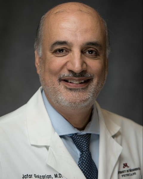
Jafar Golzarian, MD
Professor of Radiology and Surgery<br>Division Head, Vascular and Interventional Radiology
University of Minnesota
University of Minnesota
Plymouth, Minnesota, United States
Primary Author(s)
Background: n/a
Purpose/Objective: Ideal human Hepatocellular Carcinoma (HCC) animal model, especially in middle to large size, is very important for research in liver-directed therapies. As other cancer models are difficult to produce and need multiple surgeries (in addition to using immune suppression), We have developed a much simpler technique to induce HCC in rabbits by intra-arterial injection of HEPG2 cells without immune suppression or surgery with more accuracy.
Materials & Methods: 5 Adult rabbits, with identical weights were selected. After general anesthesia, the animals were placed in supine position. Arterial access was obtained via auricular artery using a 22-G needle. A 4-French guide catheter and a microcatheter were used to access the left hepatic artery. HEPG2 cells were injected into the artery. All animals underwent MRI imaging at week 2 post procedure. Liver enzyme and tumor markers were obtained weekly. All animals were euthanized 20 days after procedure.
Results: In all rabbits, MRI demonstrated the development of liver cancer in the injected lobe between 10-14 days. In addition, tumor markers and liver enzymes increased in all animals. Image-guided liver biopsy was obtained at 14 days after procedure in 2 animals. The biopsy confirmed the development of HCC.
Conclusion: Intra-arterial injection of HEPG2 cells in the hepatic artery using auricular artery is a promising less invasive technique allowing the development of HCC.
